

Gallbladder inflammation (cholecystitis) is a potentially serious digestive condition that occurs when the gallbladder becomes irritated, swollen, or infected, most commonly due to gallstones blocking bile flow. The most critical takeaway for every patient is that persistent, severe abdominal pain requires prompt clinical evaluation to prevent tissue damage or perforation, and definitive surgical removal is the standard, safest curative approach once recurrent or acute symptoms arise.
Where Is the Gallbladder and What Does It Do?
The gallbladder (مرارة) is a small, pear-shaped digestive organ situated beneath the liver in the upper right quadrant of the abdomen (موقع المرارة في الجانب الأيمن العلوي من البطن تحت الكبد). Its primary function is to store and concentrate bile—a digestive fluid produced continuously by the liver—and release it into the small intestine through bile ducts to facilitate the breakdown and digestion of dietary fats.
Symptoms of Gallbladder Inflammation
Symptoms of cholecystitis range from episodic post-meal discomfort to incapacitating, continuous pain, typically presenting as:
- Severe Upper Right Abdominal Pain: Intense, steady ache in the upper right abdomen or epigastric region, frequently radiating to the right shoulder blade or upper back, typically worsening 30 to 90 minutes after eating fatty meals.
- Nausea and Vomiting: Recurrent bouts of nausea, bilious vomiting, and loss of appetite triggered by gallbladder contractions against a blocked outlet.
- Abdominal Tenderness: Noticeable pain and muscle guarding when the upper right abdomen is pressed, frequently accompanied by an inability to inhale deeply while the doctor palpates beneath the right rib cage (Murphy’s sign).
- Bloating and Indigestion: Chronic dyspepsia, persistent fullness, and flatulence.
Acute vs. Chronic Cholecystitis
- Acute Cholecystitis (التهاب المرارة الحاد): Develops rapidly, producing severe, unyielding pain lasting several hours or days, accompanied by fever, chills, and elevated inflammatory markers requiring immediate hospital assessment.
- Chronic Cholecystitis (التهاب المرارة المزمن): Manifests as recurrent, low-grade attacks of biliary colic (pain that waxes and wanes over weeks or months), leading to a scarred, thickened, and dysfunctional gallbladder wall.
Presentation in Women and During Pregnancy
Gallbladder disease occurs significantly more frequently in women due to estrogen and progesterone, which elevate cholesterol concentrations in bile and reduce gallbladder contractility. During pregnancy, elevated hormone levels increase the tendency for biliary stasis and stone formation; persistent right upper quadrant pain during pregnancy must never be dismissed as routine morning sickness or heartburn and warrants careful ultrasound evaluation.
Warning Signs: When to Go to the Emergency Room
Seek immediate emergency medical care if you experience any of the following “red flag” symptoms:
- Abdominal pain so severe that you cannot sit still, stand upright, or find a comfortable position.
- Pain lasting continuously for more than 4 to 6 hours without easing.
- Spiking fever, rigors, or uncontrollable chills (indicating active systemic infection).
- Noticeable yellowing of the skin or whites of the eyes (jaundice), accompanied by dark, tea-colored urine or pale, clay-colored stools (signaling a stone blocking the common bile duct).
- Inconsolable vomiting with an inability to keep oral fluids down.
Causes of Gallbladder Inflammation (أسباب التهاب المرارة)
Inflammation develops when bile becomes trapped, accumulates, and triggers an inflammatory and infectious cascade:
- Calculous Cholecystitis (التهاب المرارة الحصوي): Accounts for over 90–95% of cases. A gallstone slips into the neck of the gallbladder or the cystic duct, obstructing bile outflow, which causes pressure buildup, chemical irritation, and secondary bacterial infection.
- Acalculous Cholecystitis (التهاب المرارة اللاحصوي): Inflammation occurring without the presence of stones, accounting for roughly 5–10% of cases. It primarily arises in critically ill individuals, patients recovering from major non-biliary surgeries, severe burns, prolonged intravenous feeding (TPN), or microvascular ischemic conditions.
How It Is Diagnosed
Accurate diagnosis is fast, non-invasive, and relies on direct clinical and imaging modalities:
- Abdominal Ultrasound (سونار البطن): The primary and most sensitive diagnostic tool. It visualizes gallstones, assesses gallbladder wall thickening (a hallmark of inflammation), checks for surrounding fluid accumulation (pericholecystic fluid), and detects sonographic Murphy’s sign.
- Laboratory Blood Panels: Complete blood count (CBC) to detect elevated white blood cell counts (infection), liver function tests (LFTs) to check bilirubin and transaminases for common bile duct involvement, and pancreatic enzyme levels (amylase/lipase) to rule out concurrent pancreatitis.
- Advanced Imaging (When Needed): An abdominal CT scan or MRCP (magnetic resonance cholangiopancreatography) is utilized if ultrasound views are obstructed, or if there is clinical suspicion of a stone displaced into the bile duct.
Treatment: What a Surgeon Recommends
When addressing gallbladder inflammation (علاج التهاب المرارة), clinical recommendations depend on the presentation, but surgery represents the only curative path.
Conservative Management & Treatment Duration (مدة علاج التهاب المرارة)
Temporary non-surgical management—involving hospital admission, fasting to rest the digestive tract, IV hydration, analgesics, and intravenous broad-spectrum antibiotics—is used to calm acute infection and stabilize the patient. However, conservative measures do not eliminate the underlying stones or restore normal gallbladder function. Over 30% of patients managed purely conservatively experience another acute, often more dangerous, flare-up within months.
When Surgery Is Advised and Why Timing Matters
Surgical removal (cholecystectomy) is advised as early as possible once acute cholecystitis is confirmed—ideally within the first 72 hours of symptom onset (early laparoscopic cholecystectomy). Intervening during this window prevents dense fibrous adhesions, tissue gangrene, and duct injuries, leading to shorter hospitalizations and faster recovery compared to delaying surgery for weeks.
Laparoscopic Gallbladder Removal
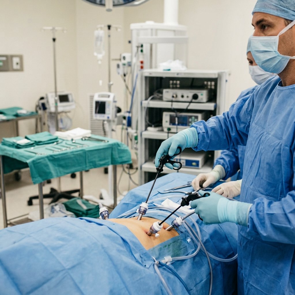
The definitive gold standard is minimally invasive laparoscopic cholecystectomy, performed through 3 or 4 tiny incisions (ranging from 5 to 10 mm) under general anesthesia. With magnified video monitoring, the cystic duct and artery are safely clipped and divided, and the diseased gallbladder is extracted cleanly with minimal pain and negligible blood loss. Learn more about the procedure, anesthesia, and technique on our dedicated Laparoscopic Gallbladder Removal Page.
Medicines: What They Can and Cannot Do
- What Medicines CAN Do: Intravenous and oral antibiotics suppress active bacterial proliferation and control sepsis; anti-inflammatory analgesics relieve acute biliary colic pain; and antispasmodics ease smooth muscle spasms.
- What Medicines CANNOT Do: Medications cannot dissolve calcified gallstones effectively, cannot repair a chronically scarred and thickened gallbladder wall, and cannot prevent recurrent blockages or complications once stones have formed. Relying on medication as a permanent substitute for surgery in symptomatic gallbladder disease is clinically inadvisable.
Is Gallbladder Inflammation Dangerous? (هل التهاب المرارة خطير)
Yes, unattended or neglected cholecystitis is a serious, potentially life-threatening condition that carries major surgical risks:
- Gangrene and Necrosis (موت الأنسجة): Continuous distension and poor blood flow lead to tissue death in the gallbladder wall.
- Gallbladder Perforation and Peritonitis (انفجار المرارة): The weakened wall can rupture, spilling infected bile and pus into the peritoneal cavity, precipitating life-threatening sepsis.
- Empyema of the Gallbladder: The gallbladder cavity fills entirely with purulent abscess fluid under extreme pressure.
- Gallstone Pancreatitis: A migrating stone slips out of the cystic duct and blocks the pancreatic duct, triggering acute, severe inflammation of the pancreas.
- Choledocholithiasis: Obstruction of the common bile duct, causing ascending cholangitis and deep jaundice.
After Treatment: Recovery and Life Without a Gallbladder
Following laparoscopic cholecystectomy, the human body adapts seamlessly; bile continues to be produced by the liver and trickles directly into the small intestine to digest food. The vast majority of patients return home within 24 hours and resume normal desk work and light routines within 5 to 7 days without permanent dietary restrictions. For detailed postoperative instructions and phase-by-phase convalescence protocols, visit our Gallbladder Surgery Recovery Guide.
When to See a Surgeon and How to Book
If you suffer from recurrent right upper quadrant abdominal discomfort, intolerance to fatty meals, or confirmed gallstones on an ultrasound, do not wait for an acute emergency to occur. Schedule a comprehensive consultation with Dr. Ahmed Abdelsalam to evaluate your gallbladder health, review your imaging, and plan a safe, elective laparoscopic intervention.
- Practice Locations:
- Mohandessin Clinic: Central surgical consultation suite.
- Sheikh Zayed Clinic: Consultations and follow-ups for West Cairo and 6th of October.
- New Cairo Clinic: Dedicated consultation clinic for East Cairo and the Fifth Settlement.
- Booking & Inquiries (Phone / WhatsApp): +201000606733 · WhatsApp
Frequently Asked Questions
هل التهاب المرارة خطير؟ (Is gallbladder inflammation dangerous?)
Yes, if left untreated. While mild early episodes may subside temporarily, unmanaged acute inflammation can rapidly advance to gallbladder gangrene, perforation (انفجار المرارة), internal abscesses, or life-threatening abdominal infections (peritonitis). Early surgical evaluation prevents these severe complications.
اين تقع المرارة وما سبب ألمها؟ (Where is the gallbladder located and what causes its pain?)
The gallbladder is located in the upper right quadrant of your abdomen, nestled underneath the liver. Its pain is caused when a gallstone lodges in the exit duct, preventing bile outflow and causing the muscular wall to cramp and inflame against the obstruction.
Can gallstones be dissolved with medications or herbs instead of surgery?
No. Oral medications to dissolve stones are largely ineffective for typical calcified gallstones, take years to show minimal effect in rare selected cholesterol-only stones, and carry a recurrence rate exceeding 50% once stopped. Surgical removal of the diseased gallbladder remains the only definitive, permanent cure.
Can I live a normal life without a gallbladder?
Yes, entirely. The gallbladder stores bile, but it does not produce it; the liver continues to produce bile, which flows directly into the intestine to digest meals normally. After an initial healing period of a few weeks, patients enjoy an unrestricted, normal diet.
How long does gallbladder surgery take, and when can I return to work?
A laparoscopic gallbladder removal typically takes between 30 and 60 minutes. Under our modern Fast Track surgical protocols, patients are mobilized within hours, discharged from the hospital the following morning, and can return to light desk work within 5 to 7 days.
Medically reviewed by Dr. Ahmed Abdelsalam on October 17, 2026.
Related pages
Back to: Gallbladder Surgery